
Successful surgical management of large conjunctival nevus in a 10-year-old child with resection and amniotic membrane transplantation
Abstract
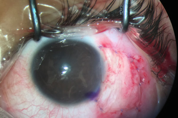
The purpose of this paper is to illustrate a case of large conjunctival nevus in a 10-year-old boy which was successfully treated with surgical excision and amniotic membrane transplant (AMT) reconstruction. The conjunctival nevus was initially noticed by the parents 1 year prior to presentation; they reported it had increased in size over the past 3 months. Slit-lamp examination revealed a pigmented conjunctival nevus measuring 5.5 mm vertically and 6.5 mm horizontally, with well-demarcated margins and presence of an intralesional cyst at the temporal bulbar conjunctiva, involving the limbus and encroaching onto the cornea. Complete resection of the conjunctival lesion and bulbar conjunctival reconstruction were performed. The histopathological examination showed conjunctival nevus. The wound healed well with vision of 6/6 and no recurrence. Surgical resection combined with AMT is a successful and an effective way to treat conjunctival
nevus.
References
Shields CL, Shields JA. Conjunctival Tumors in Children. Curr Opin Ophthalmol. 2007;18(5):351–360.
Davis JS. Skin transplantation. Johns Hopkins Hosp Rep. 1910;15:307–396.
Artur Zembowicz, Rajni V. Mandal, Pitipol Choopong. Melanocytic Lesions of the Conjunctiva. Arch Pathol Lab Med. 2010;134:1785-1792.
Khan L, Malukani M, Saxena A. Conjunctival Lesions: When Should We Perform Biopsy? Nepal J Ophthalmol. 2017;9(2):160-169.
Goktas SE, Katircioglu Y, Celik T, Ornek F. Surgical Amniotic Membrane Transplantation After Conjunctival and Limbal Tumor Excision. Arq Bras Oftalmol. 2017;80(4):242-246.
Malak TM, Bell SC. Differential expression of the integrin subunits in human fetal membranes. J Reprod Fertil. 1994;102(2):269-276.
Ahmad MS, Frank GS, Hink EM, Palestine AG, Gregory DG, McCourt EA. Amniotic Membrane Transplants in the Pediatric Population. J AAPOS. 2017;21(3):215-218.
Rock T, Bosmuller HC, Bartz-Schmidt KU, Rock D. Surgical Management of a Conjunctival Nevus with Amniotic Membrane Transplantation. Int Med Case Rep J. 2018;11:161-165.
Copyright (c) 2020 Kiew Ing Tiong, Shiivaa Manjare A/P Birapadian

This work is licensed under a Creative Commons Attribution 4.0 International License.
Authors who publish with this journal agree to the following terms:
- Authors retain copyright and grant the journal right of first publication, with the work twelve (12) months after publication simultaneously licensed under a Creative Commons Attribution License that allows others to share the work with an acknowledgement of the work's authorship and initial publication in this journal.
- Authors are able to enter into separate, additional contractual arrangements for the non-exclusive distribution of the journal's published version of the work (e.g., post it to an institutional repository or publish it in a book), with an acknowledgement of its initial publication in this journal.
- Authors are permitted and encouraged to post their work online (e.g., in institutional repositories or on their website) prior to and during the submission process, as it can lead to productive exchanges, as well as earlier and greater citation of published work (See The Effect of Open Access).
