
Multiple phakomatoses and primary open-angle glaucoma in one individual
Abstract
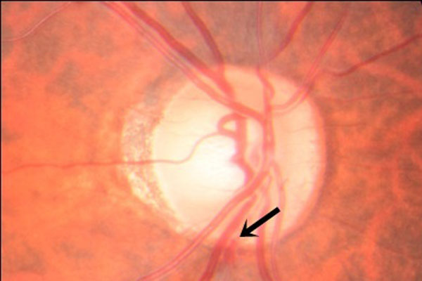
A 50-year-old woman presented with conjunctival melanosis, scleral pigmentation, and Lisch nodules in her left eye. Intraocular pressure was 24 mmHg in the right eye and 14 mmHg in the left eye. She had open angles on gonioscopy. Fundus examination showed a cup-to-disc ratio of 0.7 in the right eye, with an inferior notch and a splinter hemorrhage, and 0.6 in the left eye, with a deep cup with sloping rims. Humphrey visual fields showed an evolving superior arcuate scotoma in her right eye; the left eye was normal. Systemic examination showed axillary freckling. The patient had a family history of neurofibromatosis type 1 (NF-1), her father having been diagnosed with the condition. She had hyperpigmentation of the skin over the forehead and periocular skin on the left side. These unique ocular and systemic features were suggestive of two phakomatoses, NF-1 and nevus of Ota, in one eye, and primary open-angle glaucoma (POAG) in the other eye. that is, three pathologies present together in the same individual, which is an extremely rare occurrence.
References
Ota M. Naevus Fusco-caerunleus ophthalmomaxillaries. Tokyo Med J. 1939;63:1243–1245.
Stanford DG, Georgouras KE. Dermal melanocytosis: a clinical spectrum. Aust J Dermatol. 1996;37:19–25.
Ito M. Studies on melanin. XXII: Naevus Fusco-caeruleus acromiodeltoideus. Tohoku J Exp Med.1954;60:10-18.
Hidano A, Kajima H, Ikeda S, Mizutani, Miyasato H, Niimura M. Natural history of Naevus of Ota. Arch Dermatol.1967;95:187-195.
Teekhasaenee C, Ritch R, Rutnin U. Leelawongs N. Ocular findings in oculodermal melanocytosis. Arch Ophthalmol. 1990;108:1114-1120.
Sugar HS. Glaucoma with trabecular melanocytosis. Ann Ophthalmol.1982;14:374-375.
Kinori M, Hodgson N, Zeid JL. Ophthalmic manifestations in neurofibromatosis type 1. Surv Ophthalmol. 2018;63(4):518-533.
Huson S, Jones D, Beck L. Ophthalmic manifestations of neurofibromatosis. Br J Ophthalmol.1987;71:235–238.
Nichols JC, Amato JE, Chung SM. Characteristics of Lisch nodules in patients with neurofibromatosis type1. J Pediatr Ophthalmol Strabismus. 2003;40:293-296
Kharrat W, Dureau P, Edelson C, Caputo G. Iris mammillations: three case reports. J. Fr. Ophtalmol. 2006;29:413–417.
Ragge NK, Falk RE, Cohen WE, Murphree AL. Images of Lisch nodules across the spectrum. Eye (Lond). 1993;7:95–101.
Ceuterick SD, Van Den Ende JJ, Smets RM. Clinical and genetic significance of unilateral Lisch nodules. Bull. Soc. Belge Ophtalmol. 2005;295:49–53.
Lal G, Leavitt JA, Lindor NM, Mahr MA. Unilateral Lisch nodules in the absence of other features of neurofibromatosis 1. Am J Ophthalmol. 2003;135:567-568.
Adams EG, Stewart KMA, Borges OA, Darling T. Multiple, Unilateral Lisch Nodules in the Absence of Other Manifestations of Neurofibromatosis Type 1. Case Rep Ophthalmol Med. 2011;2011:854784. doi: 10.1155/2011/854784.
Neurofibromatosis. Conference statement. National Institutes of Health Consensus Development Conference. Arch Neurol. 1988;45:575-578.
Abdolrahimzadeh S, Plateroti AM, Recupero SM, Lambiase A. An Update on the Ophthalmologic Features in the Phakomatoses. J Ophthalmol. 2016:3043026. Epub: 2016 Jul 17.
Jain G, Jain VK, Sharma IK, Sharma R, Saraswat N. Neurofibromatosis Type 1 Presenting with Ophthalmic Features: A Case Series. J Clin Diagn Res. 2016;10(11):SR01-SR03. Epub: 2016 Nov 1.
Grant WM, Walton DS. Distinctive gonioscopic findings in glaucoma due to neurofibromatosis. Arch Ophthalmol. 1968;79:127–134.
Morales J, Imtiaz AC, Bosley TM. Glaucoma and globe enlargement associated with neurofibromatosis type1. Ophthalmology. 2009;116:1725–1730.
Croxatto JO, Charles DE, Malbran ES. Neurofibromatosis associated with nevus of Ota and choroidal melanoma. Am J Ophthalmol. 1981;92:578-80.
Gupta A, Ram J, Jain IS. Nevus of Ota associated with neurofibromatosis. Ann Ophthalmol. 1986;18:154-155.
Madke B, Kar S, Gangane N, Singh N. Phacomatosis Cesioflammea in Association With von Recklinghausen Disease (Neurofibromatosis Type I). Cutis. 2017;99(2):E35-E37.
Sangawe JL. Unilateral glaucoma. Trop Geogr Med. 1986;38(1):70-72.
Yoo YJ, Lee EJ, Kim T-W. Intereye difference in the microstructure of parapapillary atrophy in unilateral primary open-angle glaucoma. Invest Ophthalmol Vis Sci. 2016;57:4187–4193.
Zangalli CS, Ahmed OM, Waisbourd M, et al. Segmental Analysis of Macular Layers in Patients with Unilateral Primary Open-Angle Glaucoma. J Glaucoma. 2016;25:401-407.
Copyright (c) 2020 Arjit Mitra, Debarpita Chaudhury, Sumit Choudhury, Suchanda Sar, Smita Ghosh

This work is licensed under a Creative Commons Attribution 4.0 International License.
Authors who publish with this journal agree to the following terms:
- Authors retain copyright and grant the journal right of first publication, with the work twelve (12) months after publication simultaneously licensed under a Creative Commons Attribution License that allows others to share the work with an acknowledgement of the work's authorship and initial publication in this journal.
- Authors are able to enter into separate, additional contractual arrangements for the non-exclusive distribution of the journal's published version of the work (e.g., post it to an institutional repository or publish it in a book), with an acknowledgement of its initial publication in this journal.
- Authors are permitted and encouraged to post their work online (e.g., in institutional repositories or on their website) prior to and during the submission process, as it can lead to productive exchanges, as well as earlier and greater citation of published work (See The Effect of Open Access).
