
Evaluation of anterior chamber volume using Pentacam and anterior segment optical coherence tomography among normal subjects
Abstract
Aim: To compare the anterior chamber volume measurements obtained with Pentacam and derived from anterior segment optical coherence tomography.
Design: Cross-sectional study.
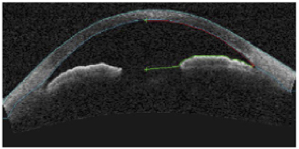
Methods: We included normal subjects who underwent a comprehensive eye examination including refraction, keratometry, Goldmann applanation tonometry, gonioscopy, anterior segment optical coherence tomography (AS-OCT) (Carl Zeiss Meditec Inc.; Dublin, CA, USA) and Pentacam (Oculus Inc.; Lynnwood, WA, USA). Fifty scans were selected for Pentacam and 12 images were selected for calculation of anterior chamber volume. Only the right eye was considered for analysis.
Results: One-hundred and nineteen eyes of 119 subjects were included for analysis. The mean age of the subjects was 42.58 ± 13.15 years, of which 74 were female and 45 were male. The mean anterior chamber volume measured using AS-OCT was 119.17 ± 26.56 mm3 and with Pentacam was 131.29 ± 34.26 mm3. The comparison of means between the two modalities was statistically significant (t = -8.857, Mean Difference (MD) = 12.11, 95% CI: (4.29, 19.95), p = 0.003). Bland-Altman plot showed poor agreement between the chamber volume measurements obtained by Pentacam and AS-OCT with MD of 12.1 mm3 (95 % CI: 41.4 to -17.1) and intra-class correlation between the two instruments was 0.94 (95% CI: 0.91, 0.96) (p < 0.0001).
Conclusion: The anterior chamber volume can be measured using Pentacam as well as AS-OCT since these measurements were reliable. However, these measurements were not interchangeable due to poor levels of agreement.
References
chamber. Invest Ophthalmol Vis Sci. 1977;16(7):633.
2. Rabsilber TM, Khoramnia R, Auffarth GU. Anterior chamber measurements using Pentacam
rotating Scheimpflug camera. J Cataract Refract Surg. 2006;32(3):456-459.
3. Wang N, Wang B, Zhai G, Lei K, Wang L, Congdon N. A method of measuring anterior chamber
volume using the anterior segment optical coherence tomographer and specialized software.
Am J Ophthalmol. 2007;143(5):879-881.
4. Yi JH, Lee H, Hong S, et al. Anterior chamber measurements by Pentacam and AS-OCT in eyes
with normal open angles. Korean J Ophthalmol. 2008;22(4):242-245.
5. Labiris G, Gkika M, Katsanos A, Fanariotis M, Alvanos E, Kozobolis V. Anterior chamber volume
measurements with Visante optical coherence tomography and Pentacam: repeatability and
level of agreement. Clin Exp Ophthalmol. 2009;37(8):772-774.
6. Fukuda S, Kawana K, Yasuno Y, Oshika T. Repeatability and reproducibility of anterior chamber
volume measurements using 3-dimensional corneal and anterior segment optical coherence
tomography. J Cataract Refract Surg. 2011;37(3):461-468.
7. Fu J, Wang X, Li S, Wu G, Wang N. Comparative study of anterior segment measurement
with Pentacam and anterior segment optical coherence tomography. Can J Ophthalmol.
2010;45(6):627-631.
8. Jonas JB, Nangia V, Gupta R, et al. Anterior chamber depth and its associations with ocular and
general parameters in adults. Clin Exp Ophthalmol. 2012;40(6):550-556.
9. Yin G, Wang YX, Zheng ZY, Yang H, Xu L, Jonas, JB. Ocular axial length and its associations in
Chinese: the Beijing Eye Study. PloS One. 2012. doi:10.1371/journal.pone.0043172.
10. Fu J, Li SN, Wang XZ, et al. Measurement of anterior chamber volume with rotating Scheimpflug
camera and anterior segment optical coherence tomography. Chin Med J. 2010;123(2):203-207.
Copyright (c) 2018 Asian Journal of Ophthalmology

This work is licensed under a Creative Commons Attribution 4.0 International License.
Authors who publish with this journal agree to the following terms:
- Authors retain copyright and grant the journal right of first publication, with the work twelve (12) months after publication simultaneously licensed under a Creative Commons Attribution License that allows others to share the work with an acknowledgement of the work's authorship and initial publication in this journal.
- Authors are able to enter into separate, additional contractual arrangements for the non-exclusive distribution of the journal's published version of the work (e.g., post it to an institutional repository or publish it in a book), with an acknowledgement of its initial publication in this journal.
- Authors are permitted and encouraged to post their work online (e.g., in institutional repositories or on their website) prior to and during the submission process, as it can lead to productive exchanges, as well as earlier and greater citation of published work (See The Effect of Open Access).
